Breast Imaging Companion (Imaging Companion Series) by Cardenosa Gilda
Author:Cardenosa, Gilda [Cardenosa, Gilda]
Language: eng
Format: epub
Publisher: Lippincot (Wolters Kluwer Health)
Published: 2011-12-26T00:00:00+00:00
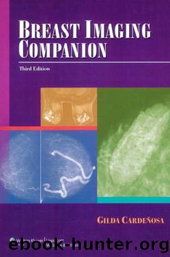
FIGURE 12-12 DCIS intermediate grade, apocrine type. Double spot compression magnification (1.8Ã) view. Punctate, round, and amorphous calcifications (arrows) of variable density demonstrate a linear orientation (segmental distribution). A few linear (casting) forms are present.
FIGURE 12-13 DCIS, intermediate to high nuclear grade. (A) Spot compression magnification (1.8Ã) view demonstrating a cluster of amorphous calcifications with low but variable density. Fibrocystic changes (e.g., hyperplasia, ADH, sclerosing adenosis, columnar alteration with prominent apical snouts and secretions), papilloma, and fibroadenoma are in the differential. (B) Specimen radiography with magnification (3.5Ã) highlights the punctate nature of amorphous calcifications seen mammographically. On a specimen, magnification can be increased so that amorphous calcifications are better âresolvedâ into tight clusters of punctate calcifications. On a patient, the amount of magnification that can be obtained is limited, and as such, tight clusters of punctate calcifications are not well defined and may appear âamorphous.â
Download
This site does not store any files on its server. We only index and link to content provided by other sites. Please contact the content providers to delete copyright contents if any and email us, we'll remove relevant links or contents immediately.
So Young, So Sad, So Listen: A Parents' Guide to Depression in Children and Young People by Philip Graham Nick Midgley(565)
Vital Signs by Izzy Lomax-Sawyers(467)
Wilderness and Survival Medicine by Ellis Chris Breen & Dr Craig(321)
Eating and Growth Disorders in Infants and Children by Joseph L. Woolston(314)
Case Studies in Adult Intensive Care Medicine by Daniele Bryden(313)
Boxed Set 1 Dermatology by Dr Miriam Kinai(288)
Manufacturing Social Distress by Robert W. Rieber(271)
Data Analysis in Sport by O'Donoghue Peter Holmes Lucy(253)
Vision and Perception by Howard Burton(236)
Yoga by Seber Isaiah(232)
Neuroradiology - Expect the Unexpected by Martina Špero & Hrvoje Vavro(228)
A History of Neuropsychology by J.Bogousslavsky & F. Boller & M.Iwata(224)
Complications in Vascular and Endovascular Surgery by Earnshaw Jonothan J;Wyatt Michael G;(215)
Basic and Advanced Laboratory Techniques in Histopathology and Cytology by Pranab Dey(214)
Manufacturing social distress : psychopathy in everyday life by Robert W. Rieber(213)
Qigong Massage for Your Child with Autism by Anita Cignolini(212)
The Psychology of Enhancing Human Performance by Gardner Frank L.;Moore Zella E.;(204)
Clinical Infectious Disease - 2020 by Weber M.D. C. G(202)
A Patient's Guide to Cataract Surgery: Normal and LASIK Reshaped Cornea by Unknown(201)
